Swine fever is a stinging disease of pigs. In the 1930s, it took place in the United States on a large scale Therefore, the disease caused pigs to have extremely high mortality. People had mistakenly thought that it was caused by a certain kind of bacteria. It was not until 1904 that it was proved that the swine fever was caused by the swine fever virus. The swine fever virus is a small RNA virus, belonging to the genus Plagioviridae and prion genus. Despite half a century of efforts, from the central government to the local departments have invested a lot of financial resources, material resources and manpower to control and eliminate swine fever, but swine fever still exists in our country, and some places are even very serious. Origin of the disease Since its first occurrence in the past more than 170 years, the disease has undergone great changes in its characteristics and epidemic forms, and this change is worldwide. First, the epidemic form has changed from a prevalent and frequent fatal epidemic and a highly lethal rate to a periodic, wave-like regional distribution. Usually a period of 3-4 years is an epidemic cycle, the epidemic points have been significantly reduced, and they have been confined to the so-called "swine stable area" in some areas and certain pig farms. Secondly, the pathological changes are not very obvious and there has been a persistent infection of swine fever virus. Third, the incidence of the characteristics of the so-called atypical chronic swine fever or mild swine fever. Significantly reduced clinical symptoms and lower mortality. Outstanding performance is the virulence syndrome of pregnant sows (that is, after the sow is infected with the swine fever virus, the virus infects the fetus through the placenta of the sow, resulting in stillbirth) and the piglet swine fever (the fetus passing through the placenta and infected with the swine fever virus is not dead, appears after birth Newborn piglet congenital tremors, the so-called "shaking disease". These "dizzly" piglets either died very quickly or became subclinically infected pigs that had long-term detoxification and transmitted the same group of pigs. For the same group of other “healthy†pigs, the same strain will have different results due to individual differences and immune status. Some pigs die from acute swine fever, while others show hidden infections and continue detoxification. Infect other pigs and pollute the environment, creating a vicious cycle. Because of the conservativeness of the antigenicity of swine fever virus, it is currently considered that there is still only one serotype. However, under the "pressure" of vaccine immunization, virulence variants of swine fever virus have emerged in many places. The term "virulence" refers to the degree of morbidity of a susceptible animal caused by a swine fever virus strain. The same strain showed different clinical symptoms due to different genetic background, age, nutritional status and immune activity of susceptible animals. Acute swine fever: The diseased pigs first showed an increase in body temperature of 40-42 °C. They were depressed, cold, sleepless, and often squeezed together or drilled into haystacks. At the beginning of the disease, constipation is more common and dry and hard stools are discharged. After 5-6 days, the sick pig developed diarrhea with a paste and water sample and mixed with blood. Conjunctivitis occurred in the affected pigs, showing an unclean mucous membrane, ulcers or bleeding spots on the gums and inner surfaces of the lips and on the tongue. In the later stage of the disease, the pigs had spot-like and patchy bleeding on the skin, and were more common in the nose, lips, ears, limbs, lower abdomen, and ventral medial skin. Bacterial infections are often secondary to late onset, and pneumonia and necrotic enteritis are more common. Chronic swine fever: mainly manifested as anemia, weight loss and general weakness. The general course of illness lasted for more than one month. The increase in body temperature was not obvious, the appetite was good or bad, and constipation and diarrhea occurred alternately. The skin of the pig's ear, tail and limbs was necrotic and even dry. Mild swine fever: The general clinical manifestations are not obvious, and the morbidity and mortality are low. Sometimes pregnant sows have miscarriage, fetal mummification, stillbirths and deformities. However, if the newborn piglet is infected with more deaths, large pigs can generally tolerate it. Pathological changes The pathological changes of swine fever are pathological changes of a septic disease. This is due to changes in edema and necrosis of vascular endothelial cells. Acute swine fever: Peripheral lymph node hemorrhage is severe and marble-like; kidney color becomes light yellow, bleeding spots are visible under the capsule, and more common in the cortex after the incision; hemorrhage in the bladder mucosa; throat, epiglottis cartilage in the upper respiratory tract Mucosal hemorrhage; The spleen is generally not enlarged, but some of the affected pigs have a round purple-red infarct on the edge of the spleen. Chronic swine fever: In addition to hemorrhagic lesions, it is characterized by necrotic enteritis. Colorectal mucosa can be seen as a round button ulcer. Mild swine fever: The main lesions of stillbirths of aborted sows are bleeding from the skin and internal organs, systemic subcutaneous edema, and pleural and abdominal effusions. Differential diagnosis Diagnosis of swine fever can be used to collect diseased pig tonsils for direct immunofluorescence antibody detection or collection of diseased swine serum. Use monoclonal antibody diagnostic reagents for enzyme linked immunosorbent assay to detect virulent swine fever virus antibodies. In addition, clinical manifestations of swine erysipelas, swine pneumoconiosis, piglet paratyphoid and swine streptococci are very similar to swine fever. Therefore, the clinical manifestations are for reference only and must be determined by comprehensive judgments based on pathological changes and epidemiology. 1. Swine erysipelas: Compared with swine fever, the infection is slow in pigs and the incidence rate is not high. The natural pores of swine erysipelas pigs have no obvious inflammation and are relatively clean, but they often die suddenly. The duration of the disease is short. The splenomegaly can be seen by necropsy. Renal stagnation and hematoma were enlarged. The lymph nodes were not marbled, and there was no significant change in the large intestine. The drug treatment significantly improved or healed. 2. Pulmonary plague: It is often distributed. The sick pig has obvious acute swelling of the throat or severe pneumonia symptoms. Breathing difficulties, mouth and nose flow out of white foam, easy to distinguish with swine fever. 3, acute paratyphoid fever: the most likely misdiagnosed with swine fever, often occurs in a pig farm, 2-4 months old piglets. The necropsy shows that the changes of the intestinal tract are different from those of the swine fever. The main manifestations are the thickening of the intestine wall of the large intestine, the rough surface inflammation of the mucosa, and the appearance of “brown†necrosis. 4, septic streptococcosis: often associated with multiple arthritis, causing movement disorders, nasal mucosa and throat, congestion, bleeding, splenomegaly. Treatment Principle 1. Immediately quarantine or cull pigs or suspicious pigs. Observe the same group of pigs in situ and strictly prohibit proliferation. 2. Emergency vaccination against swine fever vaccine should be conducted immediately in the affected area or threatened area. However, strict disinfection of medical devices must be done to prevent artificial inoculation. 3, the disease of piggery equipment, bedding, manure, the remaining feed should do a good job of disinfection. Disinfection can be quickly killed by 2% sodium hydroxide solution, 20% -30% hot grass wood grey water or 5% -10% bleaching liquid. 4. Workers are strictly prohibited from stringing, especially to prevent the breeding and epidemic prevention personnel in the affected area. 5. Units equipped with anti-pigeon serum can be used for treatment and have a certain effect on early stage pigs. 6, pay attention to feed nutrition, especially the addition of vitamins and trace elements. Preventive measures Because there is no specific drug for swine fever, it can be treated. Doing the comprehensive prevention work of the disease is the most important. Specific measures include: 1. Implementing self-cultivation and self-cultivation without importing new pigs from other places. When necessary introduction, they will be introduced and purchased in areas where there is no disease, and they will be immunized in strict accordance with the procedures. Isolation and observation for more than four weeks can be put into the farm only after it is determined to be healthy. 2. The pig farms and pig houses are often well-cleaned and cleaned and sterilized regularly (with pig disinfection). 3. Do not allow non-local personnel and other animals to enter. 4. Vaccination is the most important and effective method for preventing swine fever. The commonly used vaccines are: rabbits, rabbits, rabbits, rabbits, attenuated, rabbits, rabbits, rabbits, attenuated pigs, kidney cells, and cattle, testicular tissue, cell vaccines. There are also attenuated freeze-dried vaccines for swine fever and swine erysipelas, as well as attenuated freeze-dried vaccines for swine fever, swine erysipelas, and swine flu. The use of vaccines and swine fever immunization procedures are based on the location of the pig site, the surrounding environment, the size of the farm, the immune status of the population, and the occurrence of the disease over the years. Two immunization programs are available for reference: (1) There are no outbreak pigs in this field and surrounding areas: first exemption for 20 days, second exemption for 60 days or so. Immunize once every six months (sows can also be injected once in the delivery room). Commodity pigs: First exemption for 20 days, second exemption for 60 days old. Until the slaughter is no longer immune. (2) Piglets and piglets are often present at this site and surrounding areas: Piglets are immunized before they become immunized (i.e., they are born and immunized, and then eat milk 1 hour later). Second exemption is conducted 50 days apart, and once every six months thereafter. Commercial pigs: The piglets are immunized before milk and generally do not need to be released to slaughter thereafter.
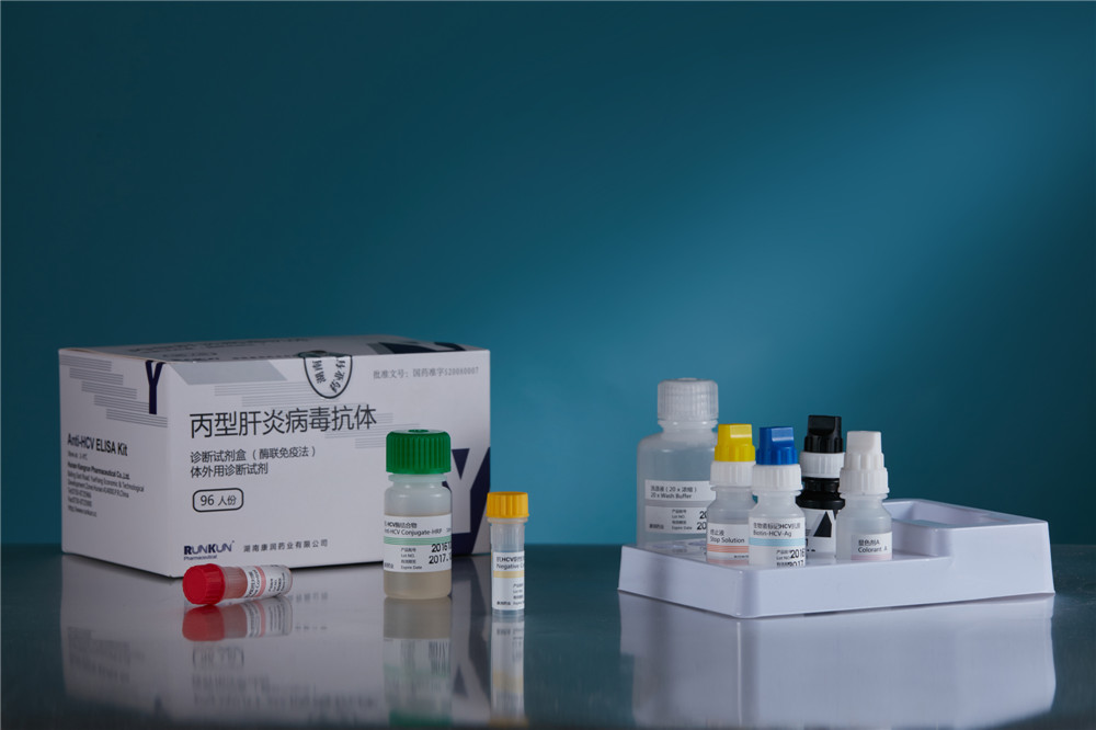
Hepatitis C Virus Antibody Diagnostic Kit
Hepatitis C virus (HCV) Antibody Diagnostic kit (enzyme-linked
immunosorbent assay, ELISA) is developed by Runkun Pharmaceutical Co.
Ltd. in cooperation with the Institute of Basic Medical Sciences under
the Academy of Military Medical Sciences of the Chinese People's
Liberation Army, and used to screen blood for HCV Antibody and to help
with the diagnosis of clinical patients. It is now China's only
diagnostic kit of double-antigen sandwich ELISA for HCV antibody
approved by the State Food and Drug Administration (SFDA). It is also
known as the diagnostic kit of "the fourth generation" for HCV antibody.

HCV Antibody
HCV Antibody,HCV Antibody Diagnostic Kit,Antibody Diagnostic Kit,HCV Diagnostic Kit
Hunan Runkun Pharmaceutical Co., Ltd. , http://www.runkun-med.com
